Tag: Vascular health

Copy of Copy of Copy of Copy of Grey Green Modern Corporate Business Presentation 11

How do you diagnose peripheral artery disease?

How Do You Diagnose Peripheral Artery Disease? Exploring Modern Techniques and Expert Insights

Peripheral Artery Disease (PAD) remains a critical health issue, affecting millions of individuals worldwide by impeding blood flow to the limbs. The journey to diagnosing this condition involves a combination of clinical evaluation, symptom assessment, and sophisticated diagnostic tools. In this article, we will delve into how medical professionals diagnose PAD, highlighting the importance of early detection and the latest advancements in medical technology.

Understanding Peripheral Artery Disease

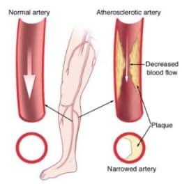
Peripheral artery disease develops when plaque builds up in the arteries that carry blood to the arms and legs, significantly reducing blood flow. Symptoms might include leg pain when walking, leg numbness or weakness, coldness in the lower leg or foot, and sores that do not heal. However, some people with PAD may not exhibit any symptoms, making diagnosis a critical challenge.

Peripheral Artery Disease (PAD) is a common circulatory problem in which narrowed arteries reduce blood flow to your limbs. When you develop PAD, your extremities — usually your legs — don’t receive enough blood flow to keep up with demand. This causes symptoms, most notably leg pain when walking (claudication).

PAD is often a sign of a more widespread accumulation of fatty deposits in your arteries (atherosclerosis). This condition may be reducing blood flow to your heart and brain, as well as your legs.

What is Peripheral Artery Disease?

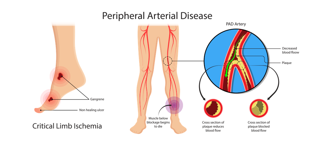
Peripheral artery disease is caused by atherosclerosis, the buildup of plaques made up of fat and cholesterol in the artery walls. As the plaques build up, the arteries become narrowed and stiffened, reducing the flow of oxygen-rich blood to the organs and other parts of the body.

Risk Factors for PAD

Understanding the risk factors for PAD is crucial because managing these can help prevent the condition or mitigate its effects if it develops. Key risk factors include:

– Age: PAD is more common in older adults, typically affecting those over 50.
– Smoking: This is a significant risk factor for PAD. Smoking contributes to the constriction and damage of arteries and raises blood pressure.
– Diabetes: High blood sugar levels associated with diabetes cause damage to blood vessels and also lead to narrowed arteries.
– High cholesterol: Excess cholesterol can contribute to plaque buildup and atherosclerosis.
– High blood pressure: This can damage blood vessels and lead to artery stiffening.
– Obesity: Being overweight is a risk factor for developing PAD.
– Sedentary lifestyle: Lack of exercise contributes to a range of conditions, including heart disease and PAD.
– Family history: A family history of PAD, heart disease, or stroke can increase risk.

Symptoms of Peripheral Artery Disease

The most common symptom of PAD is claudication, which is muscle pain or cramping in the legs or arms that is triggered by activity, such as walking, but disappears after a few minutes of rest. The location of the pain depends on the location of the clogged or narrowed artery. Calf pain is the most common location.

The severity of claudication varies widely, from mild discomfort to debilitating pain. Severe claudication can impede walking and other types of physical activity.

Other symptoms of PAD might include:

– Numbness or weakness in the legs
– Brittle toenails
– Coldness in the lower leg or foot, especially when compared with the other side
– Sores on the toes, feet, or legs that won’t heal
– A change in the color of the legs
– Hair loss or slower hair growth on the feet and legs
– Slower growth of toenails
– Shiny skin on the legs
– Erectile dysfunction, especially in men with diabetes

Complications of Peripheral Artery Disease

If left untreated, PAD can lead to severe health problems, including:

– Critical limb ischemia: This condition begins with open sores that don’t heal, an injury, or an infection of the feet or legs. Critical limb ischemia occurs when such injuries or infections progress and cause tissue death (gangrene), sometimes requiring amputation of the affected limb.
– Stroke and heart attack: The atherosclerosis that causes the signs and symptoms of PAD isn’t limited to your legs. Fat deposits also build up in arteries supplying your heart and brain.

Diagnosis and Treatment

Diagnosis of PAD begins with a physical examination. A key test in diagnosing PAD is the Ankle-Brachial Index (ABI), a simple, non-invasive test in which the blood pressure in the ankles is compared with the blood pressure in the arms. Other imaging tests, such as ultrasound, angiography, and MRI, might be used to highlight the state of the arteries.

Treatment for PAD involves lifestyle changes such as quitting smoking, exercising, and eating a healthy diet. In more severe cases, medication or surgeries such as angioplasty or bypass surgery might be necessary to open up or reroute blood vessels.

Understanding peripheral artery disease is crucial for early detection and treatment, which can significantly improve the quality of life and reduce the risk of complications such as heart attack or stroke. If you suspect you have symptoms of PAD, it is important to consult a healthcare professional for a thorough evaluation and appropriate management.
Peripheral Arterial Disease (PAD) | cdc.gov

Initial Consultation and Physical Examination

The first step in diagnosing PAD often involves a thorough medical history and physical examination. Doctors will inquire about any symptoms of leg pain, family history of cardiovascular disease, and risk factors such as smoking, diabetes, and hypertension. During the physical exam, the doctor will check for weak pulses in the legs and feet, signs of poor wound healing, and any changes in the color and temperature of the limbs.

Initial Consultation and Physical Examination for Diagnosing Peripheral Artery Disease

When suspecting Peripheral Artery Disease (PAD), the initial consultation and physical examination are crucial first steps in diagnosing the condition. These initial evaluations help healthcare providers not only identify the presence of PAD but also assess the extent and severity of the disease. This stage is vital for developing an effective management plan tailored to the patient’s specific needs.

The Importance of a Detailed Medical History

The first part of the initial consultation involves taking a detailed medical history. This is a critical component as it helps to identify any symptoms that might be indicative of PAD and any risk factors that could predispose the individual to the disease. During this phase, the healthcare provider will ask questions about:

– Symptoms: Inquiring about symptoms such as leg pain or cramps during walking or exercise, which might relieve upon resting, numbness, weakness, or any changes in the color or temperature of the legs.
-Lifestyle habits: This includes smoking history, exercise routines, and dietary habits.
– Medical history: Discussing existing conditions like diabetes, hypertension, high cholesterol, or any family history of cardiovascular disease.
– Medications: Understanding any current medications is important, as some can affect blood flow.

Conducting the Physical Examination

Following the discussion of medical history, the physical examination begins, which is pivotal in observing physical signs that suggest PAD. The examination typically includes several key assessments:

– Pulse measurement: The doctor will check for weak or absent pulses in the legs and feet. Pulse points that are routinely examined include the femoral artery in the groin, the popliteal artery behind the knee, and the dorsal pedis and posterior tibial arteries at the foot.
– Inspection of symptoms and signs: The healthcare provider will look for signs of poor circulation such as shiny skin, color changes, slower hair growth, and ulceration.
– Temperature comparison: Comparing the temperature of different parts of the legs and feet can help identify areas where blood flow might be reduced due to narrowed arteries.
– Capillary refill time: This involves pressing on the toenail or the skin to see how quickly the color returns after being blanched (turned white), which can indicate slowed blood flow.
– Ankle-brachial index (ABI): Although usually part of a more detailed diagnostic procedure, a basic ABI might be performed during the physical examination. This involves measuring blood pressure at the ankle and in the arm to compare the two as a ratio, which can indicate reduced blood flow characteristic of PAD.

What Comes After the Initial Examination?

If the history and physical exam suggest PAD, further diagnostic tests will likely be recommended. These could include more detailed blood flow studies, like duplex ultrasonography, computed tomography angiography (CTA), or magnetic resonance angiography (MRA) to visualize the blood vessels more clearly and identify specific locations and severity of blockages.

In addition to guiding further diagnostic steps, the initial consultation and physical examination help in the risk stratification of the patient. They provide essential data that can be used to tailor interventions, ranging from lifestyle modifications and medications to potentially more invasive procedures like angioplasty or surgery, depending on the severity of the disease.

Peripheral Artery Disease | The Foundation to Advance Vascular Cures

The Ankle-Brachial Index (ABI) Test

A key diagnostic test for PAD is the Ankle-Brachial Index (ABI). This simple, non-invasive test compares the blood pressure in the ankle with the blood pressure in the arm. An ABI ratio of less than 0.90 usually indicates PAD. This test is highly effective as both a screening tool and a diagnostic measure and can be performed in a doctor’s office.

The Ankle-Brachial Index (ABI) Test: A Key Diagnostic Tool for Peripheral Artery Disease

The Ankle-Brachial Index (ABI) test is a simple, non-invasive, and highly effective diagnostic tool used to detect Peripheral Artery Disease (PAD). This test compares the blood pressure in the ankle with the blood pressure in the arm to assess blood flow and identify any blockages or narrowing in the arteries. The ABI test is not only pivotal for diagnosing PAD but also serves as an indicator of the potential risk for other cardiovascular diseases.

How is the ABI Test Performed?

The ABI test is straightforward and can be performed in a healthcare provider’s office with standard blood pressure equipment and a Doppler ultrasound device. The steps involved in conducting the ABI test include:

1. Preparation: The patient is asked to lie flat and rest for about 5 to 10 minutes before the test begins, as resting blood pressure is needed for accurate measurement.
2. Measuring Arm Blood Pressure: Blood pressure cuffs are placed on both arms, and the higher of the two readings is used for the comparison.
3. Measuring Ankle Blood Pressure: The cuffs are then placed on both ankles, and a Doppler device is used to listen to the blood flow in the ankle as the cuff is inflated and then slowly deflated. The pressure at which the pulse is re-detected in the ankle as the cuff deflates is the ankle systolic pressure.
4. Calculation: The ABI is calculated by dividing the higher of the ankle pressures by the higher of the arm pressures.

Interpreting ABI Values

The results of the ABI test are given as a ratio, which can indicate normal blood flow or the presence of PAD:

– Normal ABI: An ABI ratio of 1.0 to 1.4 suggests normal blood flow without significant blockage.
– Mild to Moderate PAD: An ABI ratio of 0.91 to 0.99 may suggest the presence of mild to moderate PAD.
– Severe PAD: An ABI ratio of 0.90 or less is indicative of severe PAD, suggesting significant blockage.
– Above Normal: An ABI ratio greater than 1.4 may indicate rigid arteries and the need for further cardiovascular evaluation, often seen in patients with diabetes.

Benefits of the ABI Test

The ABI test offers several advantages in the clinical setting:

– Non-invasive and Safe: The ABI test involves no injections, no radiation, and is painless, making it a preferred first-line diagnostic tool.
– Cost-effective: Compared to other imaging tests, the ABI test is relatively inexpensive and can be performed quickly.
– Predictive Value: Besides diagnosing PAD, the ABI can indicate a higher risk of cardiovascular events such as heart attacks and strokes, especially in patients with no symptoms.

Limitations and Considerations

While the ABI test is highly valuable, it has some limitations:

– Calcification of Arteries: In patients with severely calcified arteries, such as those with long-standing diabetes, the arteries may not compress when the cuff is inflated, leading to falsely elevated ABI results.
– Operator Dependency: The accuracy of the ABI test can depend on the experience of the healthcare provider performing the test, especially in interpreting Doppler signals.

Doppler and Ultrasound Imaging

For more detailed imaging, doctors may use Doppler and ultrasound technology. These tools help visualize blood flow in the arteries and identify specific locations of blockages. By assessing the flow of blood, these tests not only confirm the diagnosis of PAD but also help in determining the severity of the disease.

Doppler and Ultrasound Imaging: Essential Tools in Diagnosing Peripheral Artery Disease

Doppler and ultrasound imaging represent critical components in the diagnostic process for Peripheral Artery Disease (PAD). These non-invasive imaging techniques provide valuable insights into the blood flow dynamics within the arteries, particularly those in the lower limbs, helping clinicians assess the presence and extent of arterial blockages or narrowing.

Understanding Doppler and Ultrasound Imaging

Ultrasound imaging uses high-frequency sound waves to create images of the inside of the body. When combined with Doppler technology, it not only shows the structure of blood vessels but also measures the movement of blood through these vessels, allowing for the assessment of both anatomical details and functional blood flow information.

How Doppler and Ultrasound Imaging Work

The procedure typically involves the following steps:

1. Preparation: The patient is positioned comfortably, usually lying down, and the area to be examined is exposed.
2. Application of Gel: A clear gel is applied to the skin over the arteries to be examined. This gel helps the ultrasound probe make secure contact with the skin and eliminates air pockets between the probe and the skin surface, which can interfere with sound waves.
3. Probe Placement: The ultrasound technician or doctor moves the probe (transducer) over the area of interest. The probe emits sound waves that bounce off the blood cells moving through the arteries and return to the probe.
4. Image and Sound Wave Analysis: The ultrasound machine processes the sound waves to create images and sounds that can be interpreted by the physician. The Doppler effect allows the device to capture changes in the frequency of the returning sound waves caused by the movement of blood through the arteries.
5. Assessment of Blood Flow: The Doppler signals provide information about the speed and direction of blood flow. Different colors on the Doppler image may indicate the speed and direction of blood flow, which helps identify areas where blood flow is restricted or blocked.

Advantages of Doppler and Ultrasound Imaging

– Non-invasive and Safe: This method does not require incisions or exposure to ionizing radiation, making it safe for repeated use.
– Real-time Results: Doppler and ultrasound imaging provide real-time pictures of blood flow and arterial structure, allowing immediate assessment.
– Detailed Visualization: These techniques can show the thickness of the arterial wall, the presence of plaques, and the severity of narrowing in the arteries.

Applications in PAD Diagnosis

Doppler and ultrasound imaging are particularly useful for:

– Locating Blockages: Identifying the specific locations of arterial blockages or stenosis.
– Assessing Severity: Evaluating the severity of the blockage and how it affects blood flow.
– Guiding Treatment: Assisting in planning medical or surgical treatments, such as angioplasty or bypass surgery.
– Monitoring Progress: Checking the effectiveness of treatments and monitoring progression of the disease over time.

Limitations

While Doppler and ultrasound imaging are highly effective, certain factors can limit their accuracy:

– Operator Skill: The quality of the imaging and the accuracy of the interpretation can depend heavily on the skill and experience of the operator.
– Physical Limitations: In patients with severe obesity or excessive gas in the intestines, obtaining clear images can be challenging.
– Deep Vessel Imaging: Imaging deep vessels or those obscured by bone can sometimes be difficult.
Peripheral Artery Disease | CTVS Texas

Computed Tomography Angiography (CTA) and Magnetic Resonance Angiography (MRA)

In cases where more detailed images are needed, Computed Tomography Angiography (CTA) and Magnetic Resonance Angiography (MRA) may be utilized. These advanced imaging techniques provide high-resolution images of the arteries, helping pinpoint where arteries are narrowed or blocked.

Treadmill Exercise Test

For patients who can tolerate physical activity, a treadmill exercise test may be recommended. This test helps assess the severity and functional impact of PAD as patients walk until they feel leg pain. Monitoring the walking distance and time helps evaluate the severity of symptoms and effectiveness of potential treatments.

Blood Tests

Blood tests may not directly diagnose PAD, but they are crucial for assessing risk factors and ruling out other conditions. Typical blood tests might include cholesterol levels, triglycerides, diabetes screening, and markers for inflammation such as C-reactive protein (CRP).

Specialist Consultations

In complex cases, or when the diagnosis is uncertain, referral to a vascular specialist may be necessary. These specialists can perform additional tests, such as angiography, where a contrast dye is injected into the arteries before X-rays are taken to view blood flow and pinpoint any blockages.

FAQs on Diagnosing Peripheral Artery Disease

What is the most common symptom of peripheral artery disease?
The most common symptom of PAD is claudication, which manifests as muscle pain or cramping in the legs or arms that occurs during activities such as walking and subsides with rest.

How reliable is the ankle-brachial index test for diagnosing PAD?
The ABI is highly reliable for diagnosing PAD in symptomatic individuals, especially those with typical symptoms like claudication. It is quick, non-invasive, and cost-effective.

Can peripheral artery disease be diagnosed at home?
While PAD cannot be diagnosed at home, individuals can monitor symptoms and risk factors. Professional medical evaluation is necessary for an accurate diagnosis.

Is PAD reversible with medication?
While PAD is not entirely reversible, medications can alleviate symptoms and stop the progression of the disease. Lifestyle changes are also crucial.

How often should someone at risk of PAD be tested?
Individuals at risk of PAD, especially smokers and those with diabetes, high blood pressure, or high cholesterol, should undergo regular screenings as recommended by their healthcare provider.

Conclusion

Diagnosing peripheral artery disease is crucial for managing the disease and improving quality of life. Through a combination of patient history, physical exams, and both basic and advanced diagnostic tests, healthcare providers can effectively identify and manage this condition. Early diagnosis and treatment can significantly reduce the risk of complications, such as heart attack, stroke, and limb amputation. Always consult with a healthcare provider for guidance and the most appropriate testing and management strategies for PAD.

DR. ALOK KUMAR UDIYA

Well-known Interventional Radiologist Dr. ALOK KUMAR UDIYA is currently a consultant at The CARE CHL, an Indore hospital with NABH accreditation. He has a distinguished medical career and has studied at numerous top federal, state, and international superspecialty medical institutes.

He earned his M.B.B.S. from M G M Medical College in Indore and then M.D. in radiodiagnosis from Lady Hardinge Medical College at Delhi University.

Following that, he completed a fellowship in neuro- and vascular interventional radiology at the Sanjay Gandhi Post Graduate Institute of Medical Sciences (SGPGIMS) in Lucknow, where he gained extensive experience in diagnostic imaging along with hepatobiliary, peripheral vascular, and neurovascular interventions.

The prestigious Institute of the liver and biliary sciences Hospital (ILBS), New Delhi, awarded him a P.D.C.C. (Post Doctoral fellowship) in Hepatobiliary intervention, where he also received further in-depth instruction and advanced training in hepatobiliary and transplant imaging and interventions.

Moreover, he completed a six-month Neuro-Interventional Fellowship (FINR) at the famous University of Zurich, where he received specialized training from Professor Anton Valavanis in the endovascular management of stroke and aneurysm. https://cvicvascular.com/

Contact Us

Contact No. –  099993 78980

Email – dralokudiya@gmail.com

Hospital

Care CHL
Address – AB Rd, near L.I.G Square, RSS Nagar, Indore, Madhya Pradesh 452008

Time – 10am to 5pm

Clinic
Address – 403 Panama Tower Geeta Bhawan Square Near Crown Palace, Indore, Madhya Pradesh
Time – 6pm To 8pm

Read More –

Carotid Stenting – https://test.mangalcart.com/carotid-stenting/

What Does Early Signs of Gangrene Look Like? – https://test.mangalcart.com/what-does-early-signs-of-gangrene-look-like/

What is the cost of AVM embolization in India? – https://test.mangalcart.com/cost-of-avm-embolization-in-india/

Green and Blue Modern Minimalist Zoom Virtual Background

Carotid Stenting : Navigating the Arteries

Introduction

In the intricate landscape of medical interventions, carotid stenting emerges as a beacon of hope for those grappling with carotid artery disease. This comprehensive guide aims to shed light on the intricacies of carotid stenting, breaking down the procedure, its significance, risks, and advancements in simple language.

Understanding Carotid Artery Disease

Before delving into carotid stenting, it’s essential to comprehend the menace it aims to tackle – carotid artery disease. This condition involves the narrowing of the carotid arteries, major blood vessels that supply blood to the brain, typically due to the accumulation of plaque. This plaque can obstruct blood flow, leading to serious consequences such as strokes or transient ischemic attacks (TIAs).

What is Carotid Stenting?

Carotid stenting is a minimally invasive procedure designed to treat carotid artery disease. It involves the insertion of a stent, a small mesh tube, into the affected carotid artery to widen it and improve blood flow to the brain. This procedure is an alternative to carotid endarterectomy, a surgical intervention with a longer recovery period.

The Procedure: Step by Step:

  1. Patient Evaluation
    Before recommending carotid stenting, thorough patient evaluation is crucial. This includes assessing the severity of carotid artery disease, the patient’s overall health, and their medical history.
  2. Angiography
    The journey begins with angiography, a diagnostic procedure that uses contrast dye and X-rays to create detailed images of the carotid arteries. This aids in determining the location and extent of blockages.
  3. Insertion of the Catheter
    A catheter is then threaded through the blood vessels to reach the site of the blockage. This is often done through the femoral artery in the groin area.
  4. Guidewire and Balloon Placement
    A guidewire is carefully maneuvered through the catheter to the site of the blockage. Subsequently, a balloon is inflated at the tip of the catheter to widen the narrowed artery, creating a space for the stent.
  5. Stent Placement
    The stent, a small mesh tube often made of metal, is then inserted and expanded at the site of the blockage. The stent acts as a scaffold, keeping the artery open and facilitating improved blood flow.
  6. Post-procedure Assessment
    Following stent placement, the catheter is removed, and the medical team evaluates the success of the procedure through additional angiography. This ensures that the stent is properly positioned, and blood flow is restored.

Benefits of Carotid Stenting

  1. Minimally Invasive
    Carotid stenting is less invasive compared to traditional carotid endarterectomy. The use of small incisions and local anesthesia contributes to a quicker recovery.
  2. Reduced Recovery Time
    Patients undergoing carotid stenting typically experience shorter hospital stays and quicker recovery times, allowing them to return to their daily lives sooner.
  3. Effective Blood Flow Restoration
    The primary goal of carotid stenting is to restore normal blood flow to the brain, reducing the risk of strokes and TIAs.

Risks and Considerations

While carotid stenting is generally considered safe, like any medical procedure, it comes with its own set of risks. Some considerations include:

  1. Risk of Stroke
    There is a slight risk of stroke during or after the procedure. This risk is carefully weighed against the potential benefits, and the medical team takes precautions to minimize it.
  2. Allergic Reactions
    Some individuals may experience allergic reactions to the contrast dye used during angiography. This risk is assessed beforehand, and alternative measures are taken if necessary.
  3. Bleeding or Infection
    As with any invasive procedure, there is a minimal risk of bleeding or infection at the catheter insertion site.
  4. Restenosis
    In some cases, the treated artery may narrow again over time, requiring further intervention. Regular follow-ups and lifestyle modifications are essential to mitigate this risk.

Advancements in Carotid Stenting

The field of interventional cardiology is dynamic, and carotid stenting is no exception. Recent advancements aim to enhance the safety and efficacy of the procedure:

  1. Embolic Protection Devices
    These devices are designed to capture and remove debris that may break loose during the procedure, reducing the risk of embolic strokes.
  2. Drug-Eluting Stents
    Similar to those used in coronary interventions, drug-eluting stents release medications to prevent restenosis, offering a longer-term solution.
  3. Patient Selection Criteria
    Improved understanding of patient risk profiles allows for more precise selection criteria, ensuring that individuals most likely to benefit from carotid stenting are identified.

Conclusion

In the realm of vascular interventions, carotid stenting stands as a beacon of progress, offering a less invasive alternative for individuals grappling with carotid artery disease. While risks are inherent, advancements in technology and a nuanced understanding of patient profiles continue to refine the procedure, enhancing its safety and efficacy.

As with any medical decision, the choice between carotid stenting and alternative treatments should be made collaboratively between the patient and their healthcare team, taking into account individual health status, preferences, and the potential risks and benefits associated with each option. In navigating the arteries, carotid stenting is a testament to the strides made in medical science to provide effective, tailored solutions for a healthier future.

DR. ALOK KUMAR UDIYA

Well-known Interventional Radiologist Dr. ALOK KUMAR UDIYA is currently a consultant at The CARE CHL, an Indore hospital with NABH accreditation. He has a distinguished medical career and has studied at numerous top federal, state, and international superspecialty medical institutes.

He earned his M.B.B.S. from M G M Medical College in Indore and then M.D. in radiodiagnosis from Lady Hardinge Medical College at Delhi University.

Following that, he completed a fellowship in neuro- and vascular interventional radiology at the Sanjay Gandhi Post Graduate Institute of Medical Sciences (SGPGIMS) in Lucknow, where he gained extensive experience in diagnostic imaging along with hepatobiliary, peripheral vascular, and neurovascular interventions.

The prestigious Institute of the liver and biliary sciences Hospital (ILBS), New Delhi, awarded him a P.D.C.C. (Post Doctoral fellowship) in Hepatobiliary intervention, where he also received further in-depth instruction and advanced training in hepatobiliary and transplant imaging and interventions.

Moreover, he completed a six-month Neuro-Interventional Fellowship (FINR) at the famous University of Zurich, where he received specialized training from Professor Anton Valavanis in the endovascular management of stroke and aneurysm.

Contact Us

Contact No. –  099993 78980

Email – dralokudiya@gmail.com

Hospital

Care CHL
Address – AB Rd, near L.I.G Square, RSS Nagar, Indore, Madhya Pradesh 452008

Time – 10am to 5pm

Clinic
Address – 403 Panama Tower Geeta Bhawan Square Near Crown Palace, Indore, Madhya Pradesh
Time – 6pm To 8pm

Read More-

Unlocking the Power of Neuro Interventions: A Comprehensive Guide – https://test.mangalcart.com/unlocking-the-power-of-neuro-interventions-a-comprehensive-guide/

What is Vascular Surgery? A Lifesaving Specialization – https://test.mangalcart.com/what-is-vascular-surgery-a-lifesaving-specialization/

Oncology Interventions: A Comprehensive Guide – https://test.mangalcart.com/oncology-interventions/

Green Food Recipe Tutorial YouTube Thumbnail

What is Vascular Surgery? A Lifesaving Specialization

Introduction

In the intricate web of our bodies, the vascular system serves as a crucial lifeline, responsible for the circulation of blood and the transportation of essential nutrients to our organs. This complex network of blood vessels, arteries, and veins can, unfortunately, become compromised due to various medical conditions, which can lead to serious health issues. Vascular surgery is a specialized branch of medicine that focuses on diagnosing and treating these conditions, ultimately saving lives and improving the quality of life for countless individuals. In this comprehensive guide, we will delve into the world of vascular surgery, exploring its importance, common procedures, and the incredible impact it has on patient well-being.

Understanding the Vascular System

Before we embark on the journey of understanding vascular surgery, it’s essential to grasp the basics of the vascular system. The vascular system consists of a vast network of blood vessels that work together to transport oxygen and nutrients throughout the body. These vessels can be divided into three main categories:

Arteries: Arteries are thick-walled blood vessels that carry oxygen-rich blood away from the heart to the various tissues and organs in the body.

Veins: Veins are responsible for returning oxygen-depleted blood from the body’s tissues back to the heart, where it can be oxygenated once again.

Capillaries: Capillaries are the smallest blood vessels that connect arteries and veins. They enable the exchange of oxygen, nutrients, and waste products between blood and body tissues.

The vascular system plays a pivotal role in maintaining the body’s overall health, and any dysfunction or damage to this system can have serious consequences.

The Importance of Vascular Surgery

Vascular surgery is a medical specialty dedicated to the diagnosis, treatment, and prevention of diseases and disorders that affect the blood vessels. This field of medicine is essential for several reasons:

Life-saving Interventions: Vascular surgeons are often called upon to perform life-saving procedures. Conditions like aortic aneurysms, peripheral artery disease, and deep vein thrombosis can lead to life-threatening situations, and timely intervention is crucial.

Improved Quality of Life: Vascular surgery can significantly enhance a patient’s quality of life. Procedures to alleviate symptoms of conditions like varicose veins or chronic venous insufficiency can make a substantial difference in a patient’s well-being.

Prevention of Complications: Vascular surgeons are skilled at diagnosing and treating conditions in their early stages, preventing the development of severe complications. This proactive approach is vital in reducing mortality and morbidity rates.

Common Vascular Conditions

Vascular surgeons encounter a wide range of conditions, each with its own set of symptoms, causes, and treatment options. Let’s explore some of the most common vascular conditions that require surgical intervention.

Aortic Aneurysms: An aortic aneurysm occurs when there is a weakening in the walls of the aorta, the largest artery in the body. This can lead to a ballooning or bulging of the artery, which poses a significant risk of rupture, a life-threatening event.

Peripheral Artery Disease (PAD): PAD is a condition that affects the blood vessels in the limbs, most commonly the legs. It is characterized by the narrowing or blockage of arteries due to the buildup of fatty deposits, which can result in reduced blood flow to the extremities.

Carotid Artery Disease: The carotid arteries are located in the neck and supply blood to the brain. When these arteries become narrowed or blocked due to atherosclerosis, it can increase the risk of stroke.

Varicose Veins: Varicose veins are swollen, twisted veins that usually occur in the legs. They are often a result of damaged valves within the veins, causing blood to pool and the veins to enlarge.

Deep Vein Thrombosis (DVT): DVT is a condition where a blood clot forms in one of the deep veins, often in the legs. If left untreated, the clot can break loose and travel to the lungs, causing a potentially fatal condition called pulmonary embolism.

Renal Artery Stenosis: This condition involves the narrowing of the renal arteries that supply blood to the kidneys. It can lead to high blood pressure and kidney problems.

Diagnostic Techniques in Vascular Surgery

Accurate diagnosis is the first step in effectively treating vascular conditions. Vascular surgeons use various diagnostic techniques to evaluate the health of a patient’s vascular system. Some of the most common methods include:

Medical History and Physical Examination: A thorough patient history and physical examination can provide essential clues about a vascular condition. The patient’s symptoms, risk factors, and family history are all taken into account.

Non-Invasive Vascular Testing: Vascular surgeons often employ non-invasive tests to assess blood flow and vessel health.

These tests include:

  • Duplex Ultrasound: This test uses high-frequency sound waves to create images of the blood vessels, helping to identify blood flow obstructions, aneurysms, and other abnormalities.
  • Ankle-Brachial Index (ABI): ABI measures the blood pressure in the ankle and arm to assess the severity of peripheral artery disease.
  • Computed Tomography Angiography (CTA): CTA is a specialized X-ray test that provides detailed images of blood vessels, allowing for the detection of blockages, aneurysms, and other abnormalities.
  • Magnetic Resonance Angiography (MRA): MRA uses magnetic fields and radio waves to create images of blood vessels. It is particularly useful for evaluating the blood vessels in the neck and brain.

Invasive Angiography: In cases where more detailed information is required, an invasive angiography procedure may be performed. During this procedure, a contrast dye is injected into the blood vessels, and X-ray images are taken to visualize the vascular system.

Treatment Options in Vascular Surgery

Once a vascular condition is diagnosed, vascular surgeons can employ a variety of treatment options, ranging from minimally invasive procedures to open surgical interventions. The choice of treatment depends on the specific condition and its severity.

Endovascular Procedures: Endovascular procedures are minimally invasive techniques that involve accessing the blood vessels through small incisions.

Some common endovascular procedures include:

  • Angioplasty: Angioplasty involves the use of a balloon-tipped catheter to open narrowed or blocked blood vessels. It is often used to treat peripheral artery disease.
  • Stent Placement: Stents are small, mesh-like tubes that are inserted into narrowed or weakened blood vessels to help keep them open. They are frequently used in conjunction with angioplasty.
  • Embolization: In this procedure, a substance is injected into a blood vessel to block or reduce blood flow to a specific area. It is commonly used to treat aneurysms or control bleeding.
  • Thrombolysis: Thrombolysis involves the administration of medications directly into a blood clot to dissolve it. This can be used to treat deep vein thrombosis or pulmonary embolism.

Open Surgical Procedures: In some cases, open surgical procedures are necessary to repair or replace damaged blood vessels.

Common open surgeries in vascular surgery include:

  • Aortic Aneurysm Repair: This procedure involves replacing or repairing the weakened section of the aorta to prevent the risk of rupture.
  • Bypass Surgery: Bypass surgery is performed to create a detour around a blocked artery. It is often used in cases of severe peripheral artery disease or carotid artery disease.
  • Endarterectomy: Endarterectomy is the removal of plaque buildup from the inner lining of an artery. It is commonly performed to treat carotid artery disease.
  • Arteriovenous Fistula Creation: This procedure is done to create a connection between an artery and a vein, often to facilitate hemodialysis for patients with kidney failure.

Conservative Management: In some cases, vascular surgeons may recommend conservative management, which involves lifestyle modifications and medications to control symptoms and slow the progression of the disease. This approach is often used for conditions like varicose veins and chronic venous insufficiency.

Recovery and Rehabilitation

Recovery and rehabilitation play a crucial role in the success of vascular surgery. The specific recovery process can vary depending on the type of procedure performed and the patient’s overall health. Here are some key aspects of the recovery and rehabilitation process:

Hospital Stay: The length of the hospital stay will depend on the type of surgery and the patient’s individual circumstances. Some endovascular procedures may allow for shorter hospital stays or even same-day discharge, while open surgeries often require longer hospitalization.

Wound Care: Patients must follow proper wound care instructions to prevent infection and promote healing. This may include changing dressings, keeping the incision site clean, and taking prescribed medications.

Physical Activity: In the early stages of recovery, patients may be advised to limit physical activity to allow the body to heal. As healing progresses, gradual reintegration of physical activity is recommended.

Medications: Patients may be prescribed medications to manage pain, prevent blood clots, or control other specific conditions. It is essential to take these medications as prescribed.

Follow-up Appointments: Patients will have regular follow-up appointments with their vascular surgeon to monitor their progress and ensure that the surgical intervention was successful.

Lifestyle Changes: Lifestyle modifications, such as quitting smoking, maintaining a healthy diet, and managing chronic conditions like diabetes and high blood pressure, are essential for long-term vascular health.

The Future of Vascular Surgery

Vascular surgery is a dynamic field, continually evolving with advancements in medical technology and techniques. The future of vascular surgery holds promising developments, including:

Minimally Invasive Innovations: Endovascular procedures are becoming increasingly sophisticated, with the development of advanced devices and techniques. These innovations will continue to reduce the invasiveness of treatments, leading to shorter recovery times and fewer complications.

Targeted Drug Therapies: Researchers are exploring targeted drug therapies for various vascular conditions. These therapies aim to prevent or treat conditions like atherosclerosis and aneurysms more effectively.

Enhanced Imaging Technology: Improvements in imaging technology, such as 3D imaging and augmented reality, will enable vascular surgeons to better visualize and navigate the vascular system during procedures, leading to improved outcomes.

Personalized Medicine: The era of personalized medicine will allow for tailored treatment plans based on an individual’s genetics, lifestyle, and specific vascular condition.

Telemedicine in Follow-up Care: Telemedicine will play a more significant role in follow-up care, making it easier for patients to consult with their vascular surgeons and receive ongoing guidance and support.

Conclusion

Vascular surgery is a medical specialty that plays a pivotal role in preserving and improving the health of individuals with vascular conditions. From life-saving interventions to procedures that enhance the quality of life, vascular surgeons are dedicated to their patients’ well-being. As the field continues to advance, patients can expect more minimally invasive treatments, better imaging technology, and personalized care that addresses their unique needs. Vascular surgery is a testament to the remarkable progress made in the field of medicine and the relentless pursuit of better solutions for vascular health.

DR. ALOK KUMAR UDIYA

Well-known Interventional Radiologist Dr. ALOK KUMAR UDIYA is currently a consultant at The CARE CHL, an Indore hospital with NABH accreditation. He has a distinguished medical career and has studied at numerous top federal, state, and international superspecialty medical institutes.

He earned his M.B.B.S. from M G M Medical College in Indore and then M.D. in radiodiagnosis from Lady Hardinge Medical College at Delhi University.

Following that, he completed a fellowship in neuro- and vascular interventional radiology at the Sanjay Gandhi Post Graduate Institute of Medical Sciences (SGPGIMS) in Lucknow, where he gained extensive experience in diagnostic imaging along with hepatobiliary, peripheral vascular, and neurovascular interventions.

The prestigious Institute of the liver and biliary sciences Hospital (ILBS), New Delhi, awarded him a P.D.C.C. (Post Doctoral fellowship) in Hepatobiliary intervention, where he also received further in-depth instruction and advanced training in hepatobiliary and transplant imaging and interventions.

Moreover, he completed a six-month Neuro-Interventional Fellowship (FINR) at the famous University of Zurich, where he received specialized training from Professor Anton Valavanis in the endovascular management of stroke and aneurysm.

Contact Us

Contact No. –  099993 78980

Email – dralokudiya@gmail.com

Hospital

Care CHL
Address – AB Rd, near L.I.G Square, RSS Nagar, Indore, Madhya Pradesh 452008

Time – 10am to 5pm

Clinic
Address – 403 Panama Tower Geeta Bhawan Square Near Crown Palace, Indore, Madhya Pradesh
Time – 6pm To 8pm

Read More –

Understanding Gastrointestinal Interventions: A Comprehensive Guide – https://test.mangalcart.com/understanding-gastrointestinal-interventions-a-comprehensive-guide/

Unlocking the Power of Neuro Interventions: A Comprehensive Guide –https://test.mangalcart.com/unlocking-the-power-of-neuro-interventions-a-comprehensive-guide/

A Comprehensive Guide to Musculoskeletal (MSK) and Orthopedic Interventions – https://test.mangalcart.com/a-comprehensive-guide-to-musculoskeletal-msk-and-orthopedic-interventions/

Yellow Minimalist Healthy Food Blog Banner 1

A Comprehensive Guide to Musculoskeletal (MSK) and Orthopedic Interventions

Introduction

Our musculoskeletal system plays a vital role in our daily lives. From walking and running to picking up objects and performing various activities, our bones, joints, muscles, and ligaments all work together to support our bodies. However, injuries, degenerative conditions, and other musculoskeletal issues can sometimes disrupt our normal functions and cause pain. This is where musculoskeletal (MSK) and orthopedic interventions come into play. In this comprehensive guide, we’ll explore what MSK and orthopedic interventions are, the conditions they address, and the various treatment options available to help people regain their mobility and quality of life.

Understanding the Musculoskeletal System

Before we delve into musculoskeletal and orthopedic interventions, it’s essential to have a basic understanding of the musculoskeletal system itself.

What is the Musculoskeletal System?

The musculoskeletal system consists of bones, muscles, joints, ligaments, and tendons, all of which work together to provide structure, support, and mobility to the body. Here’s a brief overview of each component:

1.1.1 Bones: The human body has 206 bones that serve as the framework, protecting vital organs, and providing structure. They also store minerals, such as calcium and phosphate.

1.1.2 Muscles: There are over 600 muscles in the human body, responsible for generating movement by contracting and relaxing. Muscles are attached to bones via tendons.

1.1.3 Joints: Joints are where two or more bones meet, allowing for movement and flexibility. Common types of joints include hinge, ball-and-socket, and pivot joints.

1.1.4 Ligaments: Ligaments are tough bands of connective tissue that connect bones to one another, providing stability and preventing excessive movement.

1.1.5 Tendons: Tendons connect muscles to bones, allowing the transfer of force generated by muscle contractions to the bones, leading to movement.

Now that we have a basic understanding of the musculoskeletal system let’s move on to the conditions and issues that can affect it.

Common Musculoskeletal and Orthopedic Conditions

Osteoarthritis

Osteoarthritis is one of the most prevalent orthopedic conditions. It occurs when the protective cartilage that cushions the ends of bones wears down over time. This can lead to pain, stiffness, and decreased joint mobility, particularly in weight-bearing joints like the knees, hips, and spine.

Rheumatoid Arthritis

Rheumatoid arthritis is an autoimmune disorder that primarily affects the joints. The immune system mistakenly attacks the synovium, the lining of the membranes that surround the joints, leading to inflammation, pain, and joint damage.

Fractures

Bone fractures occur when there is a break in a bone due to trauma, falls, or excessive force. Fractures can range from simple, closed fractures to complex, open fractures requiring immediate medical attention.

Tendonitis

Tendonitis is the inflammation of a tendon, often caused by repetitive movements or overuse. It can lead to pain, swelling, and limited mobility.

Sprains and Strains

Sprains involve the stretching or tearing of ligaments, while strains involve the stretching or tearing of muscles or tendons. Both conditions can cause pain and functional impairment.

Herniated Discs

A herniated disc, also known as a slipped or ruptured disc, occurs when the soft, gel-like center of a spinal disc pushes through a tear in the outer layer. This can lead to back pain and nerve compression.

Scoliosis

Scoliosis is a curvature of the spine that can affect people of all ages. While some cases are mild and may not require treatment, severe scoliosis can lead to pain and breathing difficulties.

Carpal Tunnel Syndrome

Carpal tunnel syndrome is a condition that affects the hand and wrist. It occurs when the median nerve, which runs from the forearm to the hand, becomes compressed at the wrist, leading to numbness, tingling, and weakness in the hand.

Musculoskeletal and Orthopedic Interventions

Now that we have explored some common musculoskeletal and orthopedic conditions, let’s delve into the various interventions and treatments available to address these issues and improve patients’ quality of life.

3.1 Non-Surgical Interventions

In many cases, musculoskeletal and orthopedic conditions can be managed without surgical intervention. Non-surgical treatments include:

Physical Therapy: Physical therapists design customized exercise programs to improve strength, flexibility, and mobility. They also use techniques like manual therapy and modalities such as heat, cold, and ultrasound to manage pain and improve function.

Medications: Pain relievers, anti-inflammatories, and disease-modifying drugs may be prescribed to alleviate symptoms and slow disease progression in conditions like arthritis.

Bracing and Orthotics: Custom-made or over-the-counter braces and orthotic devices can provide support, stability, and pain relief for individuals with conditions like scoliosis, plantar fasciitis, or carpal tunnel syndrome.

Injections: Corticosteroid injections can reduce inflammation and pain in affected joints or soft tissues. Hyaluronic acid injections are used to lubricate and cushion joints in some cases.

Lifestyle Modifications: Patients are often advised to make lifestyle changes, such as maintaining a healthy weight, modifying activities, and improving ergonomics to manage their conditions effectively.

3.2 Surgical Interventions

When non-surgical treatments do not provide adequate relief or if a condition has progressed to a severe stage, surgical interventions become necessary. Let’s explore some common orthopedic surgeries and procedures.

Joint Replacement Surgery

Joint replacement surgery, such as hip or knee replacement, involves removing damaged joint surfaces and replacing them with prosthetic components. These surgeries are typically performed in cases of advanced osteoarthritis or joint injuries.

Arthroscopy

Arthroscopy is a minimally invasive surgical procedure that involves inserting a tiny camera (arthroscope) and small instruments into a joint through small incisions. It is used to diagnose and treat conditions like torn ligaments, damaged cartilage, and loose bone fragments.

Spinal Surgery

Spinal surgery encompasses various procedures, including discectomy (removal of a herniated disc), spinal fusion (joining two or more vertebrae), and laminectomy (removing part of the vertebral bone). These surgeries aim to alleviate back pain and correct spinal deformities.

Tendon and Ligament Repair

Tendon and ligament injuries may require surgical repair, especially if there is a complete tear. Procedures involve reattaching the torn tissue or reconstructing it using grafts.

Fracture Fixation

Surgical procedures for fractures involve realigning and stabilizing broken bones using metal plates, screws, pins, or rods. This allows for proper healing and minimizes the risk of long-term complications.

3.3 Emerging Technologies and Techniques

The field of musculoskeletal and orthopedic interventions is constantly evolving,

with emerging technologies and innovative techniques offering new possibilities for treatment. Here are some notable advancements:

3D Printing in Orthopedics

3D printing technology allows for the creation of custom implants, prosthetics, and surgical guides tailored to individual patient needs. This technology is revolutionizing orthopedic surgery by improving precision and shortening recovery times.

Minimally Invasive Surgery

Advances in minimally invasive techniques, such as robotic-assisted surgery, have reduced surgical trauma and recovery times for many orthopedic procedures. These technologies enhance surgical precision and improve patient outcomes.

Biologics

Biologic therapies involve using substances derived from the patient’s own body, such as platelet-rich plasma (PRP) and stem cells, to promote tissue healing and regeneration. These therapies are being explored for various orthopedic applications.

Gene Therapy

Gene therapy is an emerging field in orthopedics that aims to modify the genetic factors contributing to musculoskeletal conditions. While still in the experimental stage, it holds promise for the future.

Rehabilitation and Recovery

Rehabilitation is a crucial part of the recovery process for many musculoskeletal and orthopedic conditions. After surgical interventions, patients often require rehabilitation to regain strength, mobility, and function. Physical therapy, occupational therapy, and other rehabilitation programs are tailored to individual needs and goals. The duration of rehabilitation varies based on the type and complexity of the intervention.

Physical Therapy

Physical therapists play a significant role in post-surgery recovery. They design exercise programs and techniques to rebuild strength, improve range of motion, and alleviate pain. They also educate patients on proper body mechanics to prevent future injuries.

Occupational Therapy

Occupational therapists focus on helping patients regain the skills needed for daily living and work. They address tasks like dressing, cooking, and using tools, making it easier for individuals to resume their regular activities.

Assistive Devices

In some cases, patients may need assistive devices such as crutches, wheelchairs, or braces during their recovery. These tools provide support and facilitate mobility while healing.

Pain Management

Pain management is an integral part of recovery. Medical professionals use various strategies to manage post-operative pain, including medications, nerve blocks, and physical modalities.

Home Exercises

Patients are often given specific exercises to do at home to complement their in-clinic therapy. These exercises help maintain progress and accelerate the recovery process.

Preventing Musculoskeletal Issues

Prevention is always better than treatment when it comes to musculoskeletal issues. Here are some practical steps individuals can take to reduce the risk of developing such conditions.

Maintain a Healthy Lifestyle

Maintaining a healthy weight through diet and exercise reduces the stress on weight-bearing joints, such as the knees and hips. A balanced diet rich in calcium and vitamin D helps keep bones strong.

Exercise Regularly

Regular exercise helps improve muscle strength, joint flexibility, and overall fitness. Low-impact activities like swimming, cycling, and walking are excellent options for joint health.

Practice Good Posture and Ergonomics

Maintaining proper posture while sitting, standing, and lifting objects can significantly reduce the risk of back and neck pain. Ergonomic modifications at workstations and home environments can also promote musculoskeletal health.

Avoid Overuse and Repetitive Strain

Avoid overusing certain muscle groups and take regular breaks during repetitive activities to prevent strain and overuse injuries.

Wear Appropriate Footwear

Wearing supportive and well-fitting shoes can prevent foot and lower limb problems. Custom orthotics may be necessary for those with specific foot issues.

Warm Up and Stretch

Before engaging in physical activities, it’s essential to warm up and stretch to prepare the muscles and joints. This can reduce the risk of injury.

Stay Hydrated

Proper hydration is essential for the health of your joints, as it helps maintain the lubrication of synovial fluid within them.

Living with Musculoskeletal Conditions

Living with a musculoskeletal condition can be challenging, but with the right support and strategies, individuals can manage their conditions effectively and maintain a good quality of life.

Support Networks

Joining support groups or seeking guidance from healthcare professionals can provide emotional and practical support for managing musculoskeletal conditions.

Assistive Devices

Depending on the condition, individuals may find using assistive devices helpful in daily life. These can include mobility aids, joint braces, or adaptive tools.

Pain Management

Effective pain management strategies, including medication and physical modalities, can significantly improve the daily lives of those with musculoskeletal conditions.

Physical Activity

Staying active within the limits of one’s condition is important. It can help maintain strength and flexibility and reduce the risk of secondary issues related to inactivity.

Psychological Wellbeing

Managing chronic pain and disability can take a toll on mental health. Seeking counseling or therapy to address the psychological aspects of living with a musculoskeletal condition can be valuable.

Conclusion

Musculoskeletal and orthopedic interventions are a vital part of healthcare, addressing a wide range of conditions that affect the musculoskeletal system. These interventions, whether surgical or non-surgical, aim to alleviate pain, restore function, and improve the quality of life for individuals suffering from various musculoskeletal issues.

Understanding the musculoskeletal system, recognizing common conditions, and being aware of the available treatment options can empower individuals to take proactive steps in managing their health and preventing these issues whenever possible. Additionally, staying informed about emerging technologies and innovative techniques in the field can offer hope for improved outcomes in the future.

While musculoskeletal and orthopedic conditions can be challenging, with the right interventions and support, individuals can lead fulfilling lives and continue to engage in the activities they love. The journey to musculoskeletal health often involves a combination of medical expertise, rehabilitation, and personal commitment to maintaining a healthy and active lifestyle.

DR. ALOK KUMAR UDIYA

Well-known Interventional Radiologist Dr. ALOK KUMAR UDIYA is currently a consultant at The CARE CHL, an Indore hospital with NABH accreditation. He has a distinguished medical career and has studied at numerous top federal, state, and international superspecialty medical institutes.

He earned his M.B.B.S. from M G M Medical College in Indore and then M.D. in radiodiagnosis from Lady Hardinge Medical College at Delhi University.

Following that, he completed a fellowship in neuro- and vascular interventional radiology at the Sanjay Gandhi Post Graduate Institute of Medical Sciences (SGPGIMS) in Lucknow, where he gained extensive experience in diagnostic imaging along with hepatobiliary, peripheral vascular, and neurovascular interventions.

The prestigious Institute of the liver and biliary sciences Hospital (ILBS), New Delhi, awarded him a P.D.C.C. (Post Doctoral fellowship) in Hepatobiliary intervention, where he also received further in-depth instruction and advanced training in hepatobiliary and transplant imaging and interventions.

Moreover, he completed a six-month Neuro-Interventional Fellowship (FINR) at the famous University of Zurich, where he received specialized training from Professor Anton Valavanis in the endovascular management of stroke and aneurysm.

Contact Us

Contact No. –  099993 78980

Email – dralokudiya@gmail.com

Hospital

Care CHL
Address – AB Rd, near L.I.G Square, RSS Nagar, Indore, Madhya Pradesh 452008

Time – 10am to 5pm

Clinic
Address – 403 Panama Tower Geeta Bhawan Square Near Crown Palace, Indore, Madhya Pradesh
Time – 6pm To 8pm

Read More – https://test.mangalcart.com/understanding-gastrointestinal-interventions-a-comprehensive-guide/

Unlocking the Power of Neuro Interventions: A Comprehensive Guide – https://test.mangalcart.com/unlocking-the-power-of-neuro-interventions-a-comprehensive-guide/

Gray Yellow Modern Professional Business Strategy Presentation

Navigating Vascular Health: A Closer Look at Vascular Radiology in Indore with Dr. Alok K. Udiya

Introduction

Indore, often known for its delectable cuisine and historical landmarks, is also making strides in the field of medical science. One such area of expertise is vascular radiology, a crucial discipline for diagnosing and treating vascular diseases. In this blog, we will delve into the world of vascular radiology in Indore, with a special focus on the renowned expert in the field, Dr. Alok K. Udiya.

Understanding Vascular Radiology

Vascular radiology is a specialized branch of radiology that deals with imaging and diagnosing conditions affecting the blood vessels, such as arteries and veins. It plays a pivotal role in the early detection and management of various vascular diseases, ensuring patients receive timely and accurate care.

Dr. Alok K. Udiya: The Expert in Vascular Radiology

Dr. Alok K. Udiya is a distinguished name in the field of vascular radiology in Indore. With years of experience and a stellar reputation, he has become a trusted expert for patients seeking diagnostic and interventional procedures related to vascular health.

Key Areas of Expertise

  1. Diagnostic Imaging: Dr. Alok K. Udiya employs cutting-edge imaging techniques like ultrasound, computed tomography angiography (CTA), and magnetic resonance angiography (MRA) to visualize blood vessels. These non-invasive procedures provide detailed images, aiding in the early detection of vascular conditions.
  2. Interventional Procedures: Beyond diagnosis, Dr. Udiya is skilled in performing minimally invasive interventional procedures. These include angioplasty, stent placement, embolization, and thrombolysis. These procedures are vital for treating conditions like atherosclerosis, aneurysms, and deep vein thrombosis.
  3. Patient-Centered Care: Dr. Udiya believes in a patient-centered approach to care. He takes the time to explain procedures, answer questions, and provide emotional support to his patients. This compassionate approach ensures that patients feel comfortable throughout their journey to vascular health.

Common Vascular Conditions

Vascular radiology in Indore, led by experts like Dr. Alok K. Udiya, addresses a range of vascular conditions, including:

  1. Atherosclerosis: A buildup of plaque in the arteries, which can lead to blockages and reduced blood flow.
  2. Peripheral Artery Disease (PAD): Narrowing of the arteries in the limbs, often causing leg pain and difficulty walking.
  3. Aneurysms: Weak spots in blood vessel walls that can lead to dangerous bulges and rupture.
  4. Deep Vein Thrombosis (DVT): Blood clots in the deep veins, typically in the legs, which can be life-threatening if they travel to the lungs.
  5. Varicose Veins: Enlarged and twisted veins, often causing discomfort and cosmetic concerns.

Importance of Early Diagnosis

Vascular diseases can have severe consequences if left untreated. Early diagnosis through vascular radiology is crucial for preventing complications like heart attacks, strokes, and limb amputations. Dr. Alok K. Udiya’s expertise ensures that patients in Indore have access to prompt and accurate diagnosis.

The Road to Recovery

When vascular conditions are detected in their early stages, treatment options are more effective. Dr. Udiya’s skill in interventional procedures allows for minimally invasive treatments that often result in shorter recovery times and reduced pain for patients.

Conclusion

Vascular radiology is a vital medical specialty that plays a significant role in preserving vascular health. In Indore, Dr. Alok K. Udiya stands as a beacon of expertise in this field, offering compassionate care and cutting-edge diagnostic and interventional procedures. If you or a loved one are concerned about vascular health, don’t hesitate to reach out to Dr. Udiya for expert guidance and care. Remember, early detection and intervention can make all the difference in ensuring a healthier future.

Read More –

Understanding Varicose Veins Treatment in Indore with Dr. Alok K. Udiya – https://test.mangalcart.com/understanding-varicose-veins-treatment-in-indore-with-dr-alok-k-udiya/

Interventional Radiologists: Healing from Within with Dr. Alok K. Udiya – https://test.mangalcart.com/interventional-radiologists-healing-from-within-with-dr-alok-k-udiya/

Transforming Healthcare in Indore: A Glimpse into Interventional Radiology Procedures by Dr. Alok K. Udiya – https://test.mangalcart.com/transforming-healthcare-in-indore-a-glimpse-into-interventional-radiology-procedures-by-dr-alok-k-udiya/